| |
|
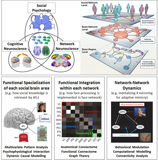
With the cutting-edge research at the intersection of social psychology, cognitive neuroscience and network neuroscience, the Wang MSN lab aims to elucidate the cognitive and brain mechanisms which allow us to interact with our social world. We take an integrative, multi-method approach, combining behavioral paradigms (motion capture, eye-tracking, psychophysiology, smartphone app), multimodal brain-imaging (functional MRI, diffusion imaging), machine learning, network analysis, and computational modeling techniques. These allow us to address research questions at multiple levels (e.g. the functional specialization of social brain regions, the functional integration of social brain networks, neural network-network interplay for adaptive social behavior, and social network dynamics in small groups).
|
|
|
|
|
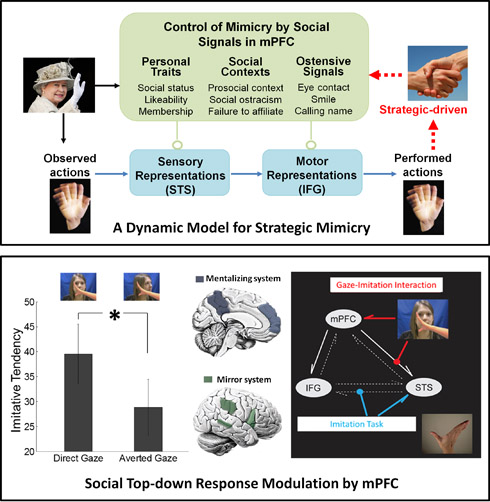
The Wang MSN lab examines the cognitive and neural foundations of human social interaction. One critical component is spontaneous mimicry. People have a tendency to unconsciously imitate other's behaviors, such as copying actions, mannerisms, facial expressions, and languages. Using motion capture, fMRI, and computational modelling, we have investigated two key questions. First, what is the purpose of mimicry? That is, under what circumstances do we mimic and why do we mimic to different degrees in different situations? Second, what brain mechanisms control and implement mimicry responses?
Over six independent papers, we provide strong evidence that mimicry is flexible and sensitive to social contexts and highlight that mimicry is a Machiavellian strategy for enhancing one's social standing. Social signals such as eye gaze (Wang & Hamilton, Biol.Lett., 2011; Wang & Hamilton, QJEP, 2014), status, likeability, and prosocial primes (Wang & Hamilton, PLoS One., 2012)
|
|
subtly and strategically control mimicry, in order to optimize the affiliative consequence of mimicry. Neuroimaing data demonstrated that mimicry is implemented by the mirror neuron system, but the medial prefrontal cortex (mPFC) plays a key role in the control of mimicry by social signals (Wang et al., J. Neurosci., 2011; Wang & Hamilton, SCAN, 2015). Computational modelling further suggested that mPFC constantly exerts top-down control on the mirror neuron system to on-line control mimicry and adaptively determines when and who to mimic (Wang et al., J. Neurosci., 2011). These findings reflect the strategic nature of mimicry that contributes to human competence in social interaction and provide important implications for our understanding of the causes and functions of mimicry (Wang & Hamilton, Front. Hum. Neurosci., 2012). Currently, the lab is using a similar approach to study other critical components of social interaction, such as body touch, interpersonal distance and facial expression.
|
|
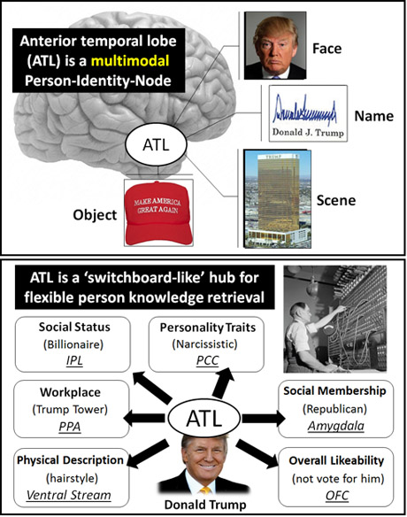
Knowledge about other people is critical for group survival. As social creatures, it is essential that we develop a rich storehouse of knowledge about other members of our social network, such as who they are, how they look and sound, where they live, and what they do for a living. Yet little is known about how and where such person knowledge is represented, stored, and retrieved in the brain.
We conducted two fMRI studies to investigate the neural dynamics when people are recalling biographies of others (Wang et al., PNAS, 2017). In study 1, we show that the anterior temporal lobe (ATL) has a function akin to a person identity node that stores abstract identity representation that is commonly embedded in multiple sources (e.g. face, name, scene, and personal object). In study 2, we found that the ATL person identity node is embedded in a neural circuit that is consistently engaged during person knowledge retrieval. MVPA analysis suggested that different pieces of person knowledge were represented in discrete nodes within this distributed person-identification circuit. Connectivity analyses further revealed that the ATL serves as a 'neural switchboard', coordinating with other nodes in a rapid and need-specific way to retrieve different contents of biographical information.
|
|
|
|
These findings endorse the ATL as a central hub for representing and retrieving person knowledge. Currently, the Wang lab is collaborating with the Olson lab at Temple University in studying the semantic and neural architecture of social relationship knowledge.
|
|
|
|
|
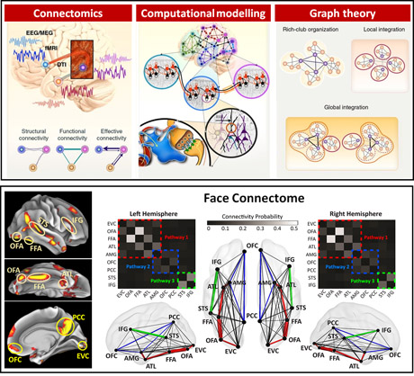
Network science and graph theory applications have recently spread widely to help in understanding how human cognitive functions are linked to neuronal network structure. This approach encompasses a variety of investigations on the node, edge, and organization of brain networks. Our past work has studied the functional specialization of the anterior temporal lobe in social knowledge network (Wang et al., PNAS, 2017), within-network functional integration of the face network (Wang et al., Nat Hum Behav, 2020) and mentalizing network (Wang et al., NeuroImage, 2021), and the network-network interaction between mirroring and mentalizing network (Wang et al., J. Neurosci., 2011).
Take the face network as an example. While much work has addressed the functional specialization of single face areas, the connectome-level organization and brain-wide mechanisms of face processing remain poorly understood.
|
|
We leveraged large-scale multimodal neuroimaging data from the Human Connectome Project Consortium to delineate the 'face connectome'. A wide range of important features were discovered, such as the network topology, core pathways, fiber composition, hemispheric lateralization, anatomy-function relations, and brain-behavior associations. This is merely the first study to systematically investigate and elucidate the 'face connectome' using such a wide variety of data (e.g. resting-state fMRI, task fMRI, diffusion imaging, and behavioral survey) and analytic approaches (e.g. graph theory, fiber analysis, machine learning, computational modelling). In the future, the Wang lab will continue to adopt this 'multimodal network approach' to elucidate the connectome-level basis of other social cognition, such as the theory of mind, imitation, empathy, social decision making, and moral cognition.
|
|
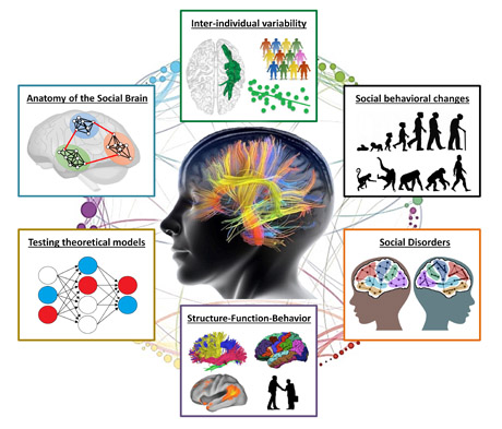
The history of human neuroscience shows an overwhelming emphasis on the functionality of gray matter, with a relative disregard of white matter. We recently strived to raise awareness of white matter functions for social cognition (Wang & Olson, TICS, 2018; Wang et al., NBR, 2018). We argue that white matter research is essential for understanding the neurobiological basis of social processes and our understanding of social cognition and social dysfunction will be incomplete until we understand the structural connectome of the social brain. Following this idea, we plan to systematically investigate how social brain areas are structurally inter-connected, which white matter structures and properties contribute to individual variability of social skills, and what roles white matter dysfunction play in social disorders.
|
|
|
|
Moreover, we are currently interested in clarifying how social brain connectome is changed during development, aging, and disease, and how this connectivity change contributes to social skill acquisition in kids and social function degradation in older adults. Due to the availability of rich open multimodal imaging resources nowadays, most of the above-mentioned questions can be addressed by working on multiple large diffusion database such as Human Connectome Project, Autism Brain Imaging Data Exchange, Philadelphia Neurodevelopmental Cohort, and Adolescent Brain Cognitive Development Study.
|
|
|
|
|
|